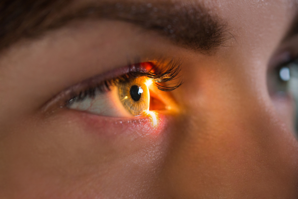
When most people schedule an eye exam, they expect to check their vision, update their prescription, and ensure their eyes are generally healthy. Traditional exams accomplish this through visual acuity tests, refraction checks, and a brief look inside the eyes - often with dilation. At Opticore Optometry Group, we use this cutting-edge tool to go beyond the basics, offering a wider, more detailed look at your retinal health without the discomfort of dilation.
Traditional Eye Exams vs. Retinal Imaging
A traditional eye exam typically includes:
- Visual acuity testing
- Refraction assessment (to determine your lens prescription)
- Eye muscle and pupil function tests
- External examination of the eye
- A basic dilated fundus exam, where the doctor uses an ophthalmoscope to view the retina
While effective for evaluating vision and some basic internal structures, this method has limitations. The field of view is relatively narrow, and certain conditions can be difficult to detect early without more advanced imaging.
Retinal imaging, on the other hand, provides a more comprehensive look inside the eye. At Opticore Optometry Group, we use the Optomap® system, a state-of-the-art retinal imaging device that captures a 200-degree widefield view of the retina in a single, non-invasive scan - compared to just 30 to 45 degrees with traditional methods.
Advantages of the Optomap® Retinal Imaging System
Optomap captures over 80% of your retina in a single image, offering a significantly broader view compared to the narrow field visible through traditional dilation and manual examination. This widefield imaging allows your optometrist to assess more of your eye’s interior in greater detail.
One of the most valuable benefits of retinal imaging is its ability to detect early signs of serious eye and systemic conditions, often before any symptoms appear. These include diabetic retinopathy, hypertension, retinal tears or detachments, macular degeneration, and glaucoma. Early detection is key to preventing vision loss and managing health proactively.
The Optomap scan is fast, non-invasive, and does not require dilation drops. That means no temporary blurred vision, no sensitivity to light, and no disruption to your day. It's an ideal solution for patients seeking a more comfortable experience during their eye exam.
Each image captured by the Optomap system is stored in your medical record, allowing for easy comparisons over time. This ongoing documentation helps your optometrist monitor for subtle changes in your retina and take action before a condition progresses.
Who Should Get Retinal Imaging?
Retinal imaging is beneficial for everyone but is especially important for patients who:
- Have diabetes or high blood pressure
- Have a family history of retinal disease or glaucoma
- Are over the age of 40
- Experience vision changes such as floaters or flashes
Even if your eyes feel fine, a retinal image can uncover silent conditions that may not yet show symptoms.
Prioritize Your Eye Health at Opticore Optometry Group
While traditional eye exams are essential for maintaining good vision, retinal imaging using the Optomap® system takes your eye care to the next level. By providing a wider, more detailed view of your retina, this advanced technology allows for earlier detection, more effective treatment, and better long-term eye health.
At Opticore Optometry Group, we’re proud to offer Optomap® retinal imaging as part of our commitment to proactive, high-quality eye care. Contact us to schedule your comprehensive eye exam and experience the difference that advanced diagnostics can make. Visit our office in Chino, Redlands, Fontana, or Riverside, California, or call (866) 202-2221 to book an appointment today.
Author: Antoinette Vu & Opticore Optometry Group


